
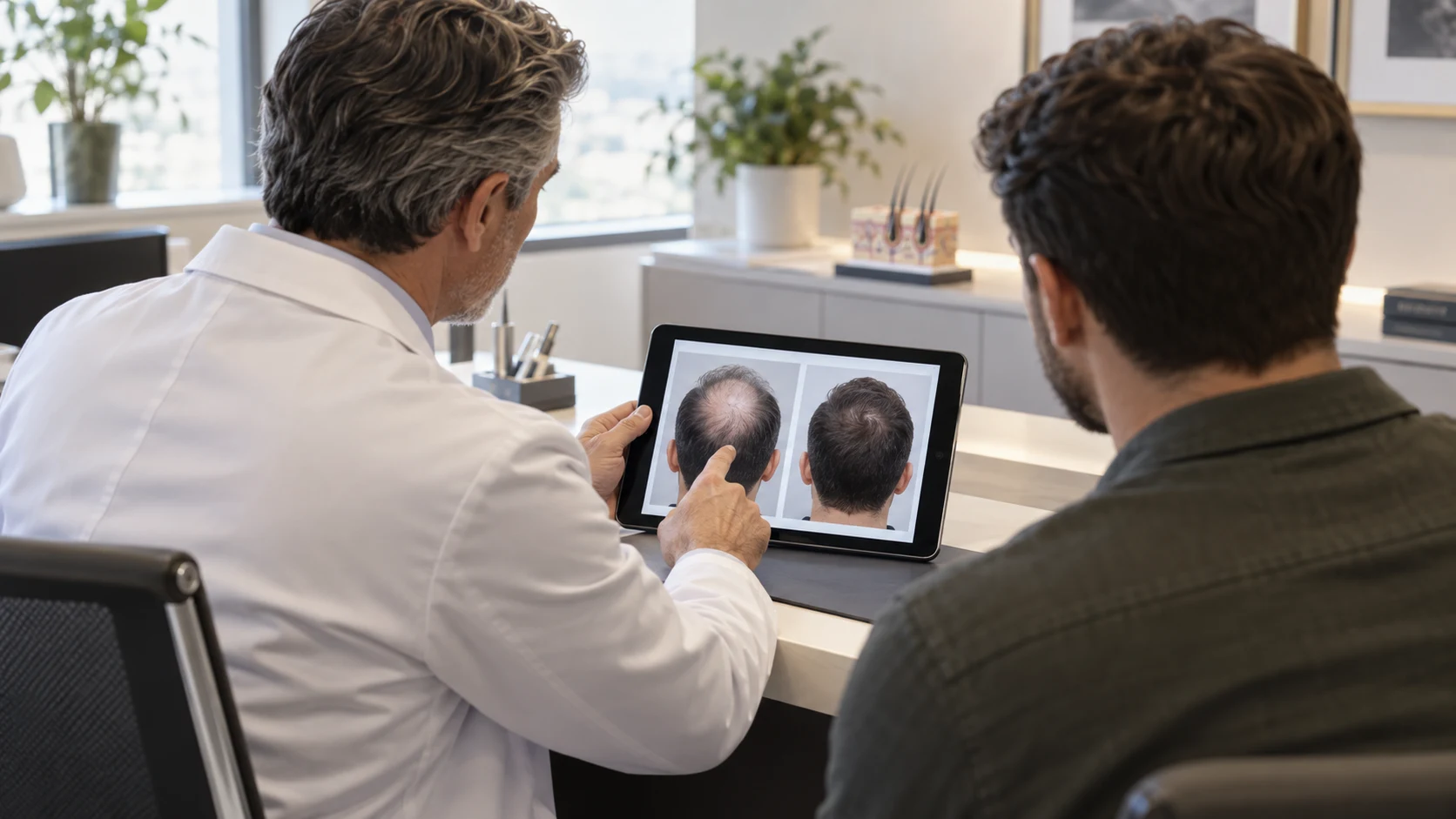
You have probably spent more hours than you would admit scrolling through other people’s scalps. Two images side by side, a number underneath, a clinic logo in the corner. And every time, the same quiet question: is that real, and could that be me?
Here is the thing nobody selling you surgery will say out loud. A before and after pair is not evidence. It is a photograph of a photograph decision — what to shoot, when to shoot it, and which pairs to publish.
The short answer: most of the visual difference between a before and after photo comes from five variables that have nothing to do with the surgery: lighting, camera angle, whether the hair is wet or dry, hair length, and how many months after the operation the second photo was taken. A trustworthy set holds all five constant. If they change between frames, you are looking at photography, not results.
The five variables that change a photo more than surgery does
Take any man with early thinning, put him under a bare overhead bulb with damp hair pushed back, and photograph him from above. Then take the same man an hour later, under soft front light, hair dry and styled forward, camera at eye level. You now have a convincing before and after. Nothing has been transplanted.
This is not a conspiracy theory. It is standard practice across a large part of the industry, and it is why experienced surgeons look at a gallery differently than patients do. They are not looking at the hair first. They are looking at the setup.
Once you know the five variables, you cannot unsee them, and you will start noticing how many published pairs quietly change three or four of them at once.
Lighting: the biggest lie in a before-and-after
Light from directly above passes through thin hair and hits the scalp, which reflects. That reflection is what makes a scalp read as bald in a photograph even when there is real coverage. Move the same light to the front and slightly lower, and the same head looks noticeably fuller.
Flash is the other culprit. A hard on-camera flash aimed at a scalp produces glare that erases fine hairs entirely, which is very convenient for a “before” image.
We wrote about this from the patient’s side in why hair looks different under different light after a transplant — the same physics that panics patients in a hotel bathroom mirror is used deliberately in marketing photography.
Angle and head tilt do half the work
Shoot down at a scalp and you see through the hair to the skin. Shoot level and you see the hair itself. A tilt of fifteen degrees changes apparent density more than a thousand grafts do.
This is why the crown is the easiest area to manipulate photographically. Almost nobody sees the top of your head from above in daily life, but that is exactly the angle chosen for the “before”.
Check the ears. If the ear position and the horizon line are different between the two frames, the camera moved, and you have lost your ability to compare anything.
Wet, dry or styled: three different heads
Wet hair clumps into separated strands and exposes scalp. Dry hair spreads, overlaps and covers. Styled hair with product covers even more. It is completely ordinary for a clinic to shoot the “before” with wet hair after a wash and the “after” dry and styled.
Look for the same state in both images. Wet-to-wet is the most honest pairing, and the least flattering, which is why it is rare.
Hair length works the same way. Longer hair lies over thinner zones and borrows density from neighbouring areas. A patient photographed at a number two clip before and with three months of growth after is showing you a haircut as much as a transplant.
When was the “after” actually taken?
This is where most galleries quietly mislead. Results at month six and results at month twelve are different pictures, and results at month four are barely a picture at all.
The realistic curve looks like this: shedding of the transplanted hairs in weeks two to six, very little visible change until month three or four, real emergence between months four and eight, and thickening that continues through month twelve and beyond. Anyone showing a dramatic after photo at three months is showing you something that will change again.
Our own breakdown of why results look different at 3, 6 and 12 months exists precisely so you can date a photo by what it shows. The crown lags further still, which is covered in crown results at 6, 8 and 12 months.
Why the graft number under the photo means less than you think
Almost every published pair carries a figure: 3,500 grafts, 4,200 grafts. It looks like data. It rarely is.
Graft counts are counted differently in different clinics, and a graft is not a hair — it is a follicular unit containing one to four hairs. Two clinics quoting the same number can be moving very different amounts of hair. The honest metric is hairs per graft and hairs per square centimetre, and almost nobody publishes it.
Graft numbers also tell you nothing about how the grafts were distributed. The same count can build a dense small hairline or a thin wide coverage. If you want the realistic ranges by pattern, how many grafts you actually need gives them without the marketing gloss.
The cases you never see
Every gallery is a survivorship record. Clinics publish their best outcomes, which is neither surprising nor dishonest by itself — no business leads with its worst day.
The problem is what the selection does to your expectations. You are comparing your realistic average outcome against a curated top decile. That comparison is unwinnable, and it is a large part of why some patients are unhappy with a technically successful result.
The useful question to ask a clinic is not “can I see your best work”. It is: can I see a case with a donor area like mine, at a similar age, photographed at twelve months, including the back of the head.
Skin tone, hair colour and contrast
Contrast between hair and scalp is a physical fact that no surgeon can change. Dark hair on pale skin shows every gap. Light or grey hair on pale skin hides them. Coarse hair covers more than fine hair at identical density.
This means a spectacular gallery result on a grey-haired patient does not predict your outcome if you have jet black hair and fair skin. It is not deception — it is simply a different optical problem.
When you look at photos, find the patient whose contrast profile matches yours. Everything else is noise.
Hair calibre works the same way and is almost never mentioned in a caption. Two patients with identical graft counts and identical surgeons can end up looking very different simply because one has thick, wiry hair and the other has fine, straight hair that lies flat against the scalp. Curl helps too: a wave lifts the hair away from the skin and covers far more area than the same number of straight hairs would. None of this is visible in a still image, which is one more reason to ask what the patient started with rather than admiring what they ended up with.
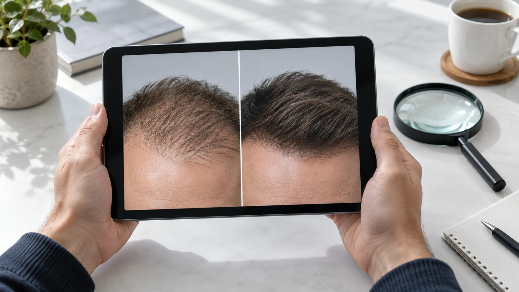
What a trustworthy before-and-after set looks like
Same room, same light, same distance, same angle, same hair state, same length where possible, with the date or month clearly stated. Multiple angles including the donor area at the back, because the donor is where honest clinics have nothing to hide and dishonest ones do.
Ideally the set includes a mid-point image at month six, not just the two endpoints. A clinic that shows the awkward middle is telling you it is not afraid of the timeline.
You can hold our own before and after gallery to exactly this standard. If a case does not show what you need, ask for another one — that request tells you a great deal about a clinic’s culture.
How to compare a photo to your own head
The mistake is comparing outcomes. The right comparison is starting points.
Find the “before” that most resembles your pattern, your age bracket and your hair characteristics, then look at what that patient got. If the gallery has no before that looks like you, the gallery cannot answer your question, however impressive it is.
If you are trying to work out whether a result like that is available to you, sending three photos of your own scalp for assessment settles it far faster than another evening of scrolling. You can request a photo assessment and get a candid answer about what your donor area can and cannot support.
Questions to ask when a clinic sends you a gallery
A short list, in the order that actually matters. How many months after surgery was the after photo taken? Were both images shot in the same room and light? How many grafts, and how many hairs per graft? What was this patient’s Norwood pattern and donor density? Can I see the back of the head? Do you have a case that failed to meet expectations, and what did you do about it?
The last question is the real test. Every surgeon has had disappointing outcomes. The ones who will discuss them openly are usually the ones worth talking to.
Our guide to the consultation questions that matter covers the rest of the conversation, and our clinic selection checklist puts the photo issue in a wider context.
Video and “unedited” clips: better, but not proof
Short videos are harder to fake than stills, because the light moves across the scalp and you can see hair behave. That makes them more useful evidence than a static pair.
They are still directed, though. Filmed in flattering light, hair pre-styled, shot from chosen angles, cropped before the awkward part. And a clip labelled “unedited” is still a clip that was selected.
Treat video as a stronger signal, not as a guarantee. The strongest signal of all is a patient you can actually speak to.
Photos taken by patients look worse — and that is normal
The photos you take of yourself at month five, in your own bathroom, under a ceiling light, will look worse than anything in any gallery. This is the single most common cause of unnecessary panic we see.
Standardise your own tracking. Same window, same time of day, same distance, dry hair, four angles, once a month. Compare month to month, never against a clinic’s marketing image.
Hair also changes appearance as it lengthens, which is why month nine can look thinner than month seven in a photograph while being objectively denser — a phenomenon explained in why results change as hair grows longer.
The “day one” photo that looks like a miracle
There is a specific image that circulates constantly: a patient photographed the morning after surgery, hairline dense and dark, scalp still dotted red. It looks like an instant result, and in a sense it is — those transplanted hairs are physically present and visible.
They will almost all fall out within six weeks. The follicle stays and regrows, but the visible shaft is shed, which is why month two often looks worse than the day after the operation. A gallery that leans on day-one and week-one images is showing you placement, not outcome.
Placement photographs are genuinely useful for a different question: they show how the surgeon designed the hairline, how irregular and natural the leading edge is, and how the angles were set. Judge design from day one. Judge density from month twelve. Confusing the two is how patients end up disappointed by a result that was always going to look like this at month three.
If a clinic only publishes day-one photographs, that is worth noticing. It usually means the twelve-month follow-up rate is low, which tells you something about how patients are handled after they fly home.
What the donor area photo tells you
The back of the head is the most informative photograph in any gallery, and the one most often missing. It is where over-harvesting shows, where scarring shows, and where the limits of a patient’s lifetime supply become visible.
What you want to see is an evenly harvested donor area with no moth-eaten patches, no visible line of dots, and no sudden change in density between the extraction zone and the untouched hair below it. Photograph it at a normal haircut length, not at three months of growth that hides everything.
A gallery with dozens of front-facing results and not a single back-of-head image is making a choice. Ask for it. A clinic that is comfortable with its extraction pattern will send it without hesitation, and one that hesitates has answered your question anyway.
This matters more than people realise if you may need a second procedure later. The donor is finite, and what a surgeon takes today shapes what is available to you in ten years.
Comparing two clinics’ galleries without fooling yourself
When you put two galleries side by side, you are usually comparing photographers rather than surgeons. The clinic with better lighting, better cameras and a consistent studio setup will look better even if the surgical work is equivalent.
Normalise before you compare. Look only at cases photographed at twelve months, only at cases with a starting pattern close to yours, and only at images where the light source appears identical in both frames. That filter typically removes most of a gallery, which is itself informative.
Then compare the things photography cannot fake: hairline irregularity, direction of growth at the temples, whether the density gradient looks natural rather than a solid block, and whether the donor is intact. Those four details survive bad lighting and expose good and bad work equally.
What you should never compare is the number of results published. Volume tells you about marketing budget, not about surgical judgement.
Why the Turkey factor makes photo literacy essential
When you are choosing a clinic in another country, photographs carry more weight than they should, because you cannot walk in, meet previous patients or see a result in person before you commit.
That is the honest reason to be more sceptical rather than less. Ask for cases with your pattern, at twelve months, with donor shots. Ask who performs which part of the operation. Our page on hair transplant in Turkey sets out what to verify before you book anything, and none of it depends on trusting a photo.
At Hairpol we would rather lose a booking to a hard question than gain one from a flattering angle.
What to do with the gallery you have been staring at
Open it again and check three things. Is the light the same in both frames? Is the after dated? Is there a case whose before looks like your head?
If all three answers are yes, the gallery is telling you something real. If any is no, it is telling you about the clinic’s photography, not about your future hairline.
And if you would rather stop guessing entirely, send your own photos for an assessment with the surgical team and ask for a case that matches your starting point. A realistic answer about your own scalp is worth more than a thousand images of someone else’s.
Find this helpful? Add Hairpol as a preferred source on Google.
Get a Free Consultation
Answer a few questions and the medical team will review your situation.
