The before-and-after photographs that clinics publish show two states: the before, and the final result. What they rarely show is everything in between — the weeks and months of biological process that separate the procedure from the outcome, each of which looks dramatically different from both the starting point and the final destination.
Understanding what a hair transplant actually looks like at each stage of recovery is one of the most practically useful things a patient can know before the procedure. It transforms the experience of recovery from a sequence of alarming surprises into a comprehensible progression — each phase making sense in context rather than prompting the anxious conclusion that something has gone wrong.
This guide goes through what the result actually looks like at each key milestone — three months, six months, and twelve months — explains why each stage looks the way it does, and gives patients an accurate framework for interpreting what they’re seeing at any point in the recovery process.
The Fundamental Principle: A Hair Transplant Doesn’t Produce Its Result Immediately
Before getting into the specific milestones, the most important conceptual shift for any patient to make is understanding that a hair transplant procedure doesn’t produce a result — it creates a biological opportunity for a result. The result itself is produced by a series of biological processes that unfold over twelve to eighteen months after the procedure.
The grafts are implanted during the procedure. Those grafts then need to establish a blood supply through revascularization, cycle through telogen after the shock of extraction and implantation, re-enter anagen and begin producing new hair, and mature through subsequent growth cycles until the hair shafts reach their full caliber and length.
Each of these stages has its own visual appearance. The appearance at month three is not a preview of the final result — it’s a snapshot of an early stage in a long biological process. The appearance at month six is different from month three and still different from the final result. The appearance at month twelve is substantially complete for most patients, though some refinement continues through month eighteen.
Patients who understand this framework interpret each milestone accurately. Those who don’t tend to panic at month three, feel cautiously hopeful at month six, and reach month twelve either pleasantly surprised or mildly disappointed — depending on whether their final result matched the inflated expectations that early-stage anxiety sometimes creates.
Immediately After the Procedure: What It Looks Like in the First Days
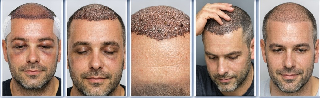
In the immediate aftermath of a hair transplant, the recipient area looks post-procedural in ways that are entirely expected but can be surprising if the patient wasn’t prepared for them.
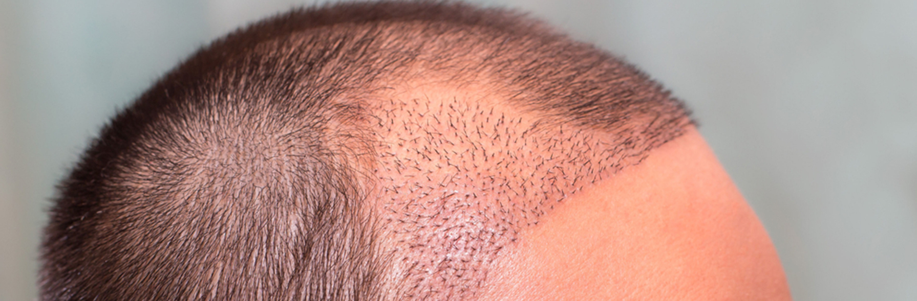
The scalp is red and slightly swollen. Small scabs or crusts form around each graft site over the first two to three days. The transplanted hairs are present — the grafts were implanted with their existing hair shafts — which creates an impression of hair in the recipient area. The donor area has the characteristic appearance of numerous tiny circular extraction sites: small red dots spread across the back and sides of the scalp.
This immediate post-procedure appearance is the most obvious-looking stage of the entire process. Most patients can’t appear in public without a hat during the first one to two weeks, and the scalp requires careful management according to the washing protocol provided by the clinic.
By days ten to fourteen, the scabs have largely shed, the donor area has healed to the point where it looks close to normal from a normal viewing distance, and the recipient area — while still slightly pink — has a more settled appearance. The transplanted hairs are still present at this stage, giving an appearance of reasonable coverage that many patients find encouraging.
This apparent density of the first two weeks is temporary and misleading as a predictor of what comes next. The transplanted hair shafts present immediately after the procedure are about to shed.
Month One: The Beginning of Shock Loss
Somewhere between weeks two and four — though the exact timing varies between patients — the shock loss phase begins. The transplanted follicles, having experienced the cumulative stress of extraction, preservation, and implantation, respond by entering telogen — the resting phase of the hair growth cycle — and shedding the hair shafts they were producing.
This is the most psychologically difficult moment in the recovery process for patients who weren’t adequately prepared for it. Hair that was there is gone. The recipient area begins looking sparse. Depending on how much native hair existed in the recipient zone before the procedure and whether that native hair is also experiencing stress-related telogen entry, the area can begin to look significantly thinner than it did before the procedure was performed.
Shock loss is not graft loss. The follicles themselves are alive and present in the scalp. The shed hairs are the existing shafts that the follicles were producing — not the follicles themselves. The follicles are entering a resting phase from which they will eventually emerge, restart their growth cycle, and produce new permanent hair in their transplanted location.
But at month one, this distinction between shock loss and graft loss offers limited visual comfort. The mirror shows thinning. The patient knows something was done. The connection between the two feels obvious even when it isn’t accurate. This is the stage that most requires intellectual understanding of the process to navigate emotionally — because the visual evidence is pointing in the wrong direction.
Month Two to Three: The Waiting Period — What the Before-and-After Galleries Never Show
Months two and three represent the stage of hair transplant recovery that before-and-after galleries systematically omit. There is no “during” photo because the during photo looks like the procedure failed.
The transplanted follicles are in telogen. They are producing no visible hair. The shock loss that began in weeks two to four has typically completed its shedding phase, and what remains is a scalp in the waiting period between the end of shedding and the beginning of new growth. This can be a period of near-complete sparseness in the recipient area — particularly for patients who had significant bald zones before the procedure.
Several specific characteristics make this period look particularly difficult:
The recipient area scalp surface, freshly healed from the procedure, reflects light differently than scalp covered by hair. Even small patches of bare scalp look more exposed and more obvious than they would later, when growing hair provides some coverage and shadow. The emotional anticipation of progress makes the absence of visible change feel more significant than objective assessment would suggest. And the overhead lighting that most patients use to assess their scalp — bathroom mirrors under ceiling lights — creates the worst possible assessment conditions, maximizing scalp visibility and minimizing any coverage the early hair might provide.
The psychological difficulty of months two and three is real and well-documented among patients who have been through the process. It is also entirely predictable and entirely temporary. The follicles that look absent at month two are working their way through telogen toward the anagen re-entry that will begin producing visible growth around months three to five.
What is actually happening during this waiting period is more encouraging than what the mirror shows. Revascularization — the growth of new blood vessels from surrounding tissue toward the implanted follicles — is progressing. The follicles are metabolically active at a maintenance level. The biological preparation for anagen re-entry is underway, entirely invisible from the outside but genuinely in progress.
Month Three: First Signs of New Growth
For most patients, new growth begins emerging somewhere around months three to five, with month three to four being a common starting point for early growth signals. This emergence is one of the most anticipated moments of the recovery process — the first visible evidence that the procedure worked and that the waiting is ending.
But what early growth looks like at month three is not what patients typically imagine. The first hairs to emerge from follicles returning from telogen are fine, often lighter in color than the mature hair these follicles will eventually produce, and unevenly distributed across the recipient area. Some zones begin showing growth while others are still entirely in telogen. The overall appearance at month three, even with early growth visible, is still significantly sparse — closer to the waiting-period appearance of month two than to the eventual result.
The unevenness of early growth is worth specific mention because it can be alarming to patients who interpret it as indicating that grafts survived better in some areas than others. While differential graft survival can occur, the uneven growth pattern of month three most commonly reflects the wave-like pattern of follicle anagen re-entry rather than differential survival — different follicles exiting telogen at different times in the same zone. Zones that look bare at month three are often simply lagging slightly behind zones that are already showing early growth, and the pattern becomes more even as more follicles complete their telogen phase over the following weeks.
The before-and-after comparison at month three is not useful. The three-month result is not a preview of the final outcome — it is a very early snapshot of a process still in its initial stages. Patients who compare their month-three appearance to the clinic’s twelve-month gallery are comparing incompatible states and setting themselves up for unnecessary distress.
Month Six: The Result Becomes Visible — But Still Not Complete
Month six is the first milestone at which something genuinely resembling a result becomes visible. For most patients, a substantial portion of the transplanted follicles have exited telogen and produced visible growth by this point. The hairline — if that was the primary treated area — is visible as a hairline. The density, while not yet at its maximum, is apparent. The shape of what the result will look like is emerging.
But the month-six result is still meaningfully different from the month-twelve result in several important ways, and patients who evaluate their result at six months as if it were final will frequently underestimate what they will eventually have.
Not all follicles have exited telogen by month six. Some follicles take longer to complete their resting phase and begin producing visible growth. Zones that look sparse at month six — particularly the crown, which has a slightly longer revascularization timeline than the frontal zone — may still be catching up on follicle activation rather than representing their final density.
Hair caliber at month six is not yet fully mature. The first hair shafts produced by follicles returning from telogen are often finer than the mature hair these follicles will eventually produce. As follicles complete their first full anagen cycle in the new location and establish more robust blood supply and tissue integration, subsequent cycles tend to produce progressively thicker shafts. The hair that looks somewhat thin at month six is often noticeably thicker and more substantial at month twelve, even from the same follicles.
Hair length at month six is relatively short. Short hair provides less coverage per follicle than longer hair that overlaps and layers to create shadow and depth. The same follicle density that looks sparse at month six — when hair is a few centimeters long — looks meaningfully fuller at month twelve when hair has reached a length that creates overlapping coverage. The improvement between six and twelve months is partly driven by more follicles activating, but also significantly by existing hairs becoming longer and providing more coverage per strand.
A useful benchmark for month six is that most patients are seeing approximately fifty to seventy percent of their eventual result. They can evaluate the hairline design, the overall direction of the result, and the distribution of coverage — but they cannot accurately evaluate the final density from this snapshot.
Month Nine: Substantial Maturity
Month nine marks a significant shift in how hair transplant results can be assessed. By this point, the vast majority of follicles have exited telogen and begun producing visible growth. Hair that emerged in months three to five has grown to a length where it provides meaningful overlapping coverage. Hair caliber is approaching its mature level for most follicles. The result at month nine represents a substantially complete picture — typically eighty to ninety percent of what the final result will be.
For many patients, month nine is the milestone at which they first feel genuinely satisfied with what they’re seeing. The density is sufficient, the hairline looks natural, the design decisions made before the procedure are fully visible and evaluable. Patients who have been navigating the anxiety of months two through five often experience a specific moment of recognition at month nine — seeing for the first time that the result is genuinely what they hoped for.
The remaining improvement between month nine and month twelve relates primarily to late-emerging follicles completing their anagen entry, continued maturation of hair caliber in follicles whose first anagen cycles produced finer initial shafts, and additional hair length growth that provides marginally more coverage. These are refinements rather than fundamental changes — the architecture of the result is established by month nine even if some finishing continues through month twelve and occasionally through month eighteen.
Month Twelve: The Result Benchmark
Twelve months is the standard benchmark for evaluating a hair transplant result, and for most patients it represents a substantially mature outcome. The great majority of transplanted follicles are producing hair. Hair caliber has reached close to its mature level for most follicles. Hair length has grown enough that the result can be assessed under a range of styling conditions, not just when worn very short.
The comparison between the pre-procedure state and the twelve-month result is the before-and-after comparison that genuinely reflects the outcome of the procedure. Before-and-after galleries that show twelve-month results under consistent conditions give an accurate representation of what the procedure produced — subject to all the caveats about photography conditions, hair characteristics, and individual variation that apply to any gallery assessment.
Several important characteristics of the twelve-month result are worth understanding specifically:
The twelve-month result is not static. The transplanted hair will continue to grow, cycle, and age normally. It will not fall out due to androgenetic processes — the donor dominance principle ensures that transplanted follicles retain their DHT resistance in their new location. But it will behave like any other hair in terms of growth cycles, styling, and aging. The twelve-month snapshot captures the result at one point in this ongoing process.
The surrounding native hair has continued to evolve. Native hair in areas adjacent to the recipient zone has continued its natural genetic progression during the twelve months of recovery. Patients who had some remaining native hair at the time of the procedure may find that this hair has continued to thin during the recovery period — which affects the overall density picture at month twelve, independent of the transplanted hair’s performance.
Some patients continue to see improvement beyond month twelve. The crown is the most common area where this applies — results in the crown consistently mature more slowly than frontal results, and some patients are still seeing meaningful improvement through month fifteen or eighteen. Patients who had crown procedures should not finalize their assessment of the crown result at twelve months if growth is still visibly progressing.
The Crown vs. The Frontal Zone: Different Timelines
One of the most clinically significant differences in recovery timelines is between the frontal zone and the crown, and it’s worth specific discussion because patients who had procedures addressing both areas may find the two zones developing on noticeably different schedules.
The frontal zone consistently shows earlier and more visually impactful growth than the crown. The frontal scalp has a more dense vascular network, which supports faster revascularization of implanted grafts and earlier anagen re-entry. Frontal hairs also grow in a relatively consistent forward direction, which means that as each hair grows longer it adds incrementally to coverage in a predictable way.
The crown’s anatomical characteristics produce a different growth pattern. The vascular network in the crown is somewhat less dense, revascularization takes slightly longer, and anagen re-entry of transplanted follicles is correspondingly delayed. The spiral whorl growth pattern of the crown means that early growth hairs radiate outward in multiple directions — each hair provides less unidirectional coverage than a frontal hair growing forward, and the overlapping coverage effect of longer hair takes more time to accumulate.
The practical consequence is that a patient at month six who has a clear, developing frontal result may still have a crown that looks largely unchanged from the waiting period. This is not differential graft survival — it is differential timing in a recovery process that genuinely runs on slightly different clocks in different zones of the scalp. Crown growth typically becomes clearly visible between months five and eight, and the full crown result typically takes until months twelve to eighteen to fully mature.
Why Before-and-After Comparisons Are Often Misleading
Before-and-after galleries are the primary tool patients use to evaluate clinics and set expectations, and they contain systematic biases that make them less informative than they appear.
The selection bias is fundamental: clinics publish their best results. A gallery of fifty excellent results assembled from thousands of patients doesn’t represent the distribution of outcomes — it represents the best-case tail of that distribution. Even a clinic with mediocre average results could assemble a compelling gallery.
The photography asymmetry compounds this. Before photos are typically taken under unflattering conditions — overhead lighting, wet or slicked-back hair, angles chosen to maximize the apparent degree of loss. After photos are taken under favorable conditions — side or natural lighting, hair styled for maximum coverage, angles chosen to showcase the result at its best. The same patient photographed under the before conditions at their twelve-month result would look significantly worse than they do in the after photograph — and photographed under the after conditions before the procedure, they would look significantly better than they do in the before photograph.
The timing of after photographs matters significantly. Most gallery after photographs are taken at twelve to eighteen months — when the result is at its best. They don’t show what the result looks like at five years if surrounding native hair has continued progressing, or what the result looks like under overhead lighting rather than the photographically favorable conditions used in the gallery.
Understanding these limitations doesn’t make before-and-after galleries useless — they contain real information about what a procedure can produce. But they should be evaluated critically: looking for cases with similar characteristics to your own, seeking results photographed under consistent rather than asymmetric lighting, looking for long-term results beyond twelve months, and understanding that what you’re seeing represents the favorable tail of outcomes under favorable conditions rather than a typical result under typical conditions.
How to Track Your Own Progress Accurately
Given the systematic biases in how patients typically assess their own recovery — overhead lighting, very short hair, daily checking that captures random variation rather than genuine trend — it’s worth being specific about how to track progress in a way that actually reflects biological reality.
Photograph at consistent intervals — monthly is reasonable — under consistent conditions. Natural outdoor light rather than overhead bathroom lighting. The same distance, the same angle, the same hair styling state. This consistency is what allows genuine comparison between timepoints rather than comparisons confounded by random variation in conditions.
Assess hair at the length you actually wear it, not at the shortest possible length. A result that looks sparse at a buzz cut length often looks meaningfully fuller at two to three centimeters and significantly fuller at four to five centimeters. The appropriate assessment condition is how you’ll actually wear the hair, not the worst-case length.
Compare across meaningful time intervals rather than daily. Hair growth is too slow to produce visible change day to day, and daily assessment primarily captures variation in lighting, styling, and mood rather than biological progress. Monthly comparison photographs reveal the genuine trend. Daily mirror checks primarily generate anxiety.
Understand what stage of the process you’re in before interpreting what you see. At month two, the expected appearance is sparse. At month six, the expected appearance is emerging but incomplete. At month twelve, the expected appearance is substantially complete. Interpreting month-two sparseness as failure because it doesn’t look like the month-twelve gallery is comparing incompatible states — the visual equivalent of judging a building by its appearance during the framing stage rather than after completion.
When the Result at Twelve Months Isn’t What You Expected
Most patients who reach month twelve with accurate expectations find that their result is within the realistic range of what was achievable for their specific hair characteristics, loss pattern, and donor supply. They may wish the density were higher or the hairline slightly different, but the result is recognizable as a genuine improvement and not as a disappointment relative to what the procedure could realistically deliver.
Patients who reach month twelve with genuine dissatisfaction — where the result is objectively below the realistic range for their situation rather than below an inflated expectation — are in a different position. Clinical assessment at this point can help distinguish between: results that are still developing and need more time; results that represent lower graft survival from the procedure that may benefit from a supplemental session; and results where the design or execution produced outcomes below what the patient’s characteristics would support.
The appropriate response to genuine underperformance at month twelve is a thorough consultation that honestly assesses what happened, what options exist, and what a realistic improvement path looks like — not an immediate additional procedure booked before the current result has fully matured.
At Hairpol, post-procedure support includes structured guidance at each key milestone of the recovery timeline — not because what patients see at month three requires clinical intervention, but because understanding what they’re seeing at month three is one of the most practically useful supports the clinic can provide. The before-and-after story is more complex than two photographs suggest, and patients who understand the full arc of the process navigate it significantly better than those who discover each stage without preparation.
The Bottom Line: What Each Milestone Actually Tells You
The hair transplant recovery timeline produces results that look dramatically different at each key milestone — not because the outcome is changing unpredictably, but because each stage reflects a specific phase in a biological process that is progressing exactly as it should.
At month three, you are seeing the early stage of anagen re-entry — fine, sparse, uneven early growth that represents the beginning of the result rather than the result itself. At month six, you are seeing a developing result — substantially visible but not yet complete, with continued improvement to come from late-emerging follicles, maturing hair caliber, and growing hair length. At month twelve, you are seeing a substantially complete result — the culmination of twelve months of biological process that was underway from day one of the procedure.
Each stage is exactly where it should be. The growth is coming. The biology is working. And the before-and-after that will eventually tell the story of your procedure will look nothing like the in-between — which is precisely why the in-between needs its own explanation.
